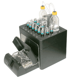
Automated pharmacology instrument
The instrument allows researchers to discover in real time the effects of their novel
drugs in living blood vessels.
It is therefore an automated pharmacology instrument with applications in University research, the Pharmaceutical Industry and Biotechnology Companies.
The instrument allows delivery of accurate concentrations of drugs to an
isolated living blood vessel contained within an organ bath that is positioned at the front
of the instrument.
The system then uses syringe pumps to accurately control pressure
and flow of fluids containing the test drugs within the blood vessel, thereby creating the
same conditions that exist in vivo. This area of research is known by a number of terms
such as “organ bath pharmacology”, “perfusion myography”, “wire myography” and
“pressure myography”.
The User obtains information on the response of the tissue at different doses of the test drug (pharmacology) which help to predict what the effects of the drug will be at later stages of clinical trials.
Biopta’s award winning patented technology aims to revolutionise in vitro pharmacology
with the introduction of our first instrument. Biopta's scientists have an international
reputation in the analysis and assessment of drug-tissue interactions in our fully GLP
human tissue CRO laboratory. These specialists in human tissue bioassays have
developed numerous pharmacological screens for many human tissues and with this
expertise they have developed the next generation of instruments for in vitro
pharmacology.
Our first instrument is an automated perfusion and wire myograph, Biopta PM-1. This
system launches our patented technology, Perf-Exion®, which uses optical properties
of the tissue to detect drug effects on a wide range of tubular tissues. Up to four
individually regulated tissues provide information on drug potency and efficacy from
sensitive measurement of changes in tissue diameter, flow, pressure and force
production.
Biopta’s unique Perf-Exion® optical technology, which allows detection of the external
and internal dimensions of tubular biological structures, has won numerous awards
including a John Logie Baird Award and Scottish Executive "SMART" and "SPUR"
awards for innovation. PM-1 is the first of Biopta’s instruments to incorporate Perf-
Exion® technology and is specially designed to capture information from isolated blood
vessels.
Perfusion myography is an in vitro "bioassay" which, by using isolated tissues that
retain their in vivo characteristics, allows dimensional analysis of tubular structures
capable of dilation and constriction. The technique has been frequently used to study
a range of blood vessels and has led to the discovery of key vascular mechanisms such
as myogenic tone and flow-mediated vasodilatation. Perfusion of blood vessels
is accepted as being more representative of normal physiology than organ bath or wire
myograph techniques, which also makes it an ideal technique for drug screening.
Sensitive measurements of inner (lumen) and outer diameter together with automated
delivery of test compounds allows concentration-response relationships to be
created that provide information on safety, efficacy and potency. Furthermore, as a
basic research tool, Biopta’s PM-1 system has revolutionised the means by which
pharmacologists can investigate the basic physiology of the cardiovascular system by
automating and de-skilling the entire process.
Perfusion studies have been particularly important in our basic understanding of the
physiology of the coronary circulation.
Given that coronary side-effects are a major concern in many classes of drugs, the
potential of an automated pharmacology system is obvious.
Among the many advantages of PM-1 is the unique ability to simultaneously capture the
responses of up to four tissues, with the pressure and flow in each vessel separately
regulated in order to mimic in vivo conditions.
The system accommodates a wide range of tissue dimensions and flow rates allowing
tubular tissues from 150 microns to 2 mm external diameter to be studied. Pressure can
be maintained with or without flow, or alternatively pressure changes can be recorded
while a constant flow rate is imposed on the vessel. Previous perfusion systems
required a high level of operator skill and time-consuming "nursing" of the instrument
through the experiment.
Biopta’s system has deskilled the tissue handling process, fluid control systems
and instrument set-up procedures. Moreover, the instrument includes a PC controlled,
user-friendly test scheduler that allows the operator to simply program the
instrument and walk away. Biopta’s system offers an unparalleled mixture of flexibility
and automation for both basic vascular research and in vitro drug screening.
Why Biopta PM-1?
The Benefits
Saves Time
Programmable experimental protocols
with 12 programmable drug vials and user friendly Windows software
Automated delivery of drugs to tissue
with pressure and flow controlled independently
Ease of tissue mounting
stainless steel cannula rather than glass with a wide range of size-matched cannula available
Saves Costs
Frees up technician time
with fully automated delivery of test compounds and programmable protocols
Quicker start up time
minimal training required with de-skilled tissue handling
Wide breadth of physiological methods
with only 1 system for wire and perfusion myography
Quality Data
Highly sensitive detection of physiological and pharmacological changes
patented Perf-Exion® technology allows measurement of the internal & external
diameter of the tissue
Improved signal to noise ratio
with accurate control of flow through syringe pumps and an intelligent pressure
feedback system
Controlled drug delivery
with precise delivery of drug concentrations
PM-1 allows four individually-regulated tissues to be studied simultaneously, yet
its modular design offers the choice of set-ups for the study of one, two, three or four tissues
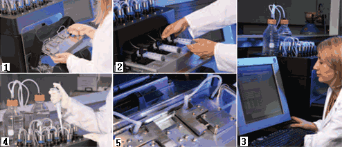
1 Ease of tissue mounting with removable tissue bath
2 Sensitive control of flow through accurate syringe pumps
3 Programmable experimental protocols with user friendly Windows software
4 Automated fluid handling with 12 programmable drug vials
5 A wide range of tissue can be studied from 150 µm to 2 mm external diameter
Technical Specification
Fluid Handling/Automation:
Vials : 12 temperature regulated vials for fully automated
drug delivery
Temperature range : 28°C – 40°C
Temperature stability : +/- 0.5°C
Main reservoir : 1 x 1000 mls
Waste reservoir : 1 x 1000 mls
Test compound vials : 10 x 15 ml vials, 2 x 60 ml vials
Pressure control : 0 – 200 mm Hg
Accuracy : +/-1 mmHg, automatic pressure maintenance with
and without flow
Flow rate : 25 µl / hr – 25 ml / min
Organ Bath:
Tissues : Suitable for a wide range of tubular tissues 150 µm to
2.0 mm external diameter
Cannula : Stainless steel
Cannula size(s) : 14 G to 35 G (2.11 mm to 0.13 mm external diameter)
Temperature range : 28°C – 40°C
Chamber filling : Automated wash & rinse programs
Software/Data Capture:
1Hz capture of : Intraluminal pressure
Internal diameter of vessel
External diameter of vessel
Longitudinal tension (perfusion)
Radial tension (wire)
Programmable : Drug additions
Flow rate
Pressure
Volume perfused
Sampling of perfusate with collection in micro-centrifuge tubes