|
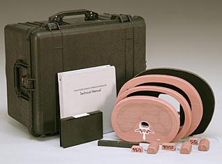
MODEL 004 INCLUDES
Tissue Equivalent lumbar section
Medium and large attenuator rings
Tissue Equivalent vertebral
inserts 50, 100, 150 mg/cc
calcium hydroxyapatite
Slice thickness gauge
Acrylic support board and
base stand
Technical manual
Graphic report software
(DOS or Windows)
Manual work sheets and
report forms (optional)
Custom foam lined carrying
case Dimensions:
22" 1/4 x 14" 1/2 x 17"
Informative patient literature
Technical hotline |
BENEFITS:
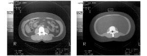
• Accurately simulates the size, shape and CT density of human tissue
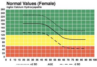
• Includes standard vertebral inserts of varying density to permit accurate correlation for quantitative studies
• Provides the age-related variable corrections for
marrow fat and mineral content
• Provides direct measure of calcium hydroxyapatite
content avoiding the need for special extrapolations
• Requires no special scanner software
• Ideal for monitoring effects of therapy on
trabecular structure
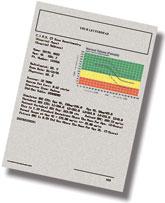
• Includes PC based report software
• Can be used immediately on all whole body
CT scanners
|
MODEL 004 computer PC software produces detailed graphic reports on your stationary.
 |
|
REFERENCES
(1 ) Levi C, Gray JE, McCullough EC, Hatery RR, The unreliability of CT numbers as absolute values. AJR 1982:139:443-447
(2) Lampmann LEH, Duursma SA, Ruys JHJ, CT densitometry in osteoporosis 1984: Martinus Nijhoff]
(3) Cann CE, Genant HK: Precise measurement of vertebral mineral content using computed tomography. J Comput Assist Tomography 4:493,1980
|