|
|
|
|
|
|
|
|
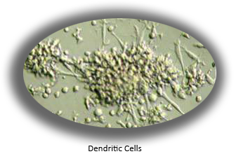
Dendritic (Langerhans) Cells

Features
- Longer Life Span in Culture
- Generated from Umbilical Cord Blood CD34+ Progenitor Cells - More Responsive Than Monocyte-Derived DC's
- Cells Expanded 400 Fold - Perform Multiple Experiments with Constant Cell Origin, Functionality
- Key Markers Present, Including CD1a, HLA-DR and Birbeck Granules
- Both Plasmacytoid DC (pDC; CD123+, CD11c-) and Myeloid DC (mDC; CD123-, CD11c+) Present
- Non-Adherent Suspension Cells - More In Vivo-Like
- Infectable with HIV-1, Transmits Virus to T-Cells
- Excellent for Allergenicity, Antigen Binding and Presentation, Viral Infection, Immuno-therapeutic (ex. "cancer vaccine") and Transfection Studies
- Dendritic Cells Integrated Into Several MatTek In Vitro Human Tissue Equivalents
- DC-Based Immunological Contract Research Available On Request (상담하기)
Applications
Dendritic Cells
- Allergenicity Testing
- Microbial Infection
- Antigen Presentation
- Vaccine Immunotherapy
- Immunotoxicity
- Induction of Tolerance
- Inflammatory / Cytokine Response
- Viral / Bacterial / Fungal Infection
- Skin Flushing Studies
- Probiotic Immunology
- Basic DC Research
Data Sheet
Dendritic cells (DC) play a key role in the immunological reactions throughout the body. Dendritic cells (DC) and their immature counterparts, Langerhans cells (LC), are highly specialized antigen-presenting cells (APC) located in the skin, mucosa, and lymphoid tissues. DC and LC play a key role in the induction phase of contact allergenicity, and it is likely that these cells can be used to develop in vitro assays for contact sensitization and other immunological reactions of the body.
The difficulty in harvesting and the short survival time of DC in culture has prevented their widespread use by researchers. Although improvements have been made, generating large number of cells has remained a limiting factor and the retention of basal (non-primed) functionality of the cells has been difficult to achieve. Finally, their response to external stimuli has often been inconsistent.
In response to this need, MatTek has developed a new method for generating DC from CD34+ progenitor cells (hematopoietic stem cells) harvested from umbilical cord blood. This improved method generates an average of >400 million DC from 1 million starting progenitor CD34+ cells.
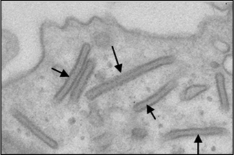
The DC express CD1a, HLA-DR, CD80, CD86, CD40, CD54, CD11c, CD206 (mannose receptor), and unlike monocyte-derived DC, the DC express BIRBECK GRANULES. The DC also express TLR-1, TLR-2, TLR-3, TLR-4, TLR-6, TLR-7 and TLR-9.
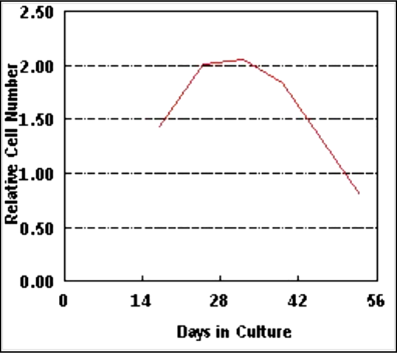
The lifespan of the DC has been increased to 29 days with detectable Birbeck granules and very small phenotypic changes over this period. The DC can be maintained for at least 14 days with no significant changes in surface marker expression or functionality.
Upon stimulation with LPS and PMA, a reproducible, significant gene and protein expression was observed; IL-12, MIP-1α, MIP-3α, IL-6, and TNF-α were upregulated. Further, the DC are capable of stimulating heterologous T cells (TC) in mixed lymphocyte reaction (MLR) and autologous TC following exposure to antigen.
The non-stimulated DC can be fractionated by FACS sorting into plasmacytoid DC (pDC; CD123+CD11c-) and myeloid DC (mDC, CD123-CD11c+). Compared to the mDC, the pDC have shorter dendrites, express lower level of CD1a (11.8% vs. 66.8%), and can be expanded 6-15 fold using specially formulated medium (mDC do not proliferate). The pDC also showed increased levels of CD86 surface expression following treatment with the allergens (n=7), but not following exposure to irritants (n=5).
Plasmacytoid Dendritic Cells are available for sale as a separate item (part number DCP-100).
The generated DC are also infectable with HIV-1 and can transmit virus to TC.
In summary, MatTek's new DC methodology overcomes many of the problems that previously plagued dendritic cell research. The DC are phenotypically similar to their in vivo counterparts, and their in vitro function in immunological reactions mimics their in vivo behavior. These cells have longer life span in culture and can be used in a broad range of immunology, virology and other functional studies.
Dendritic cells generated from umbilical cord blood were characterized for:
1) Growth curve - Using cell counts obtained at different time points (Figure 1).
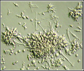
2) Morphology - using light microscopy (Figure 2),
3) Ultrastructure - using transmission electron microscopy (TEM) for the presence of the unique DC marker, Birbeck granules (Figure 3), and
4) Phenotype - using FACS analysis of surface marker expression (Table 1).
FUNCTIONAL ANALYSIS: RESPONSE TO IMMUNO-STIMULANTS
To examine the functionality of generated DC, cells were stimulated with lipopolysaccharide (LPS) or phorbol myristate acetate (PMA) as follows.
After 24 hr of exposure, culture supernatants were collected to quantify cytokines released into the culture medium using commercially available cytokine ELISA kits (Figure 4). The stimulated cells were also harvested, RNA was extracted and cytokine gene expression levels were monitored using RT-PCR (Figure 5).
As shown in Figure 4 below, culture supernatants collected from LPS or PMA stimulated cells showed elevated levels of IL-12p40, IL-8, MIP-3a, MIP-1a, and RANTES. Furthermore, as expected, DC-100 cells express message for cytokines such as IL-12p40 upon stimulation with LPS or PMA (Figure 5).

Figure 4: Cytokine production by DC-100 cells following 24 hr exposure to LPS or PMA.
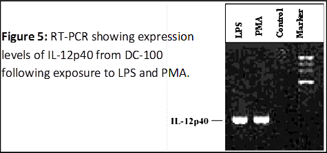
Figure 5: RT-PCR showing expression levels of IL-12p40 from DC-100 following exposure to LPS and PMA
FUNCTIONAL ANALYSIS: STIMULATION OF AUTOLOGOUS
T-CELLS
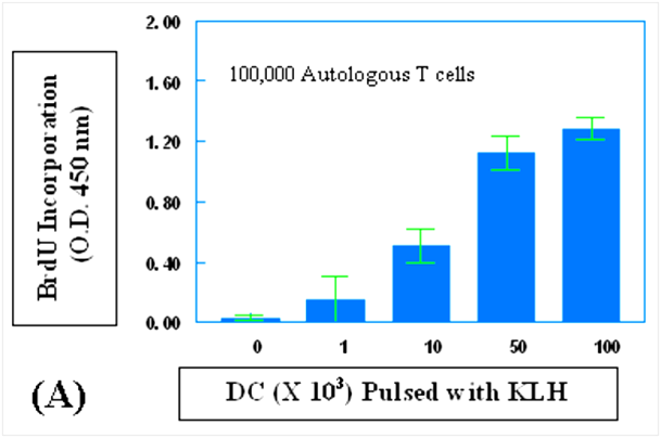 Figure 6: Proliferation of T cells (TC) induced by autologous dendritic cells (DC) pulsed with: (A) keyhole limpet hemocyanin (KLH) or (B) tetanus toxoid. DC were pulsed with either KLH (0.5 μg/ml) or tetanus toxoid (5.0 μg/ml) for one hour, treated with mitomycin C (50 μg/ml) for 30 min, and washed 2X with phosphate buffered saline. Varying numbers of the DC were then used to stimulate a fixed number of TC (100,000). The DC/TC mixture was cultured for 96 hr and then incubated for an additional 24 hr in the presence of BrdU. TC proliferation was determined by the level of BrdU incorporation as determined by the optical density (OD) at 450 nm. When no DC were added, the BrdU incorporation OD was 0.030. Figure 6: Proliferation of T cells (TC) induced by autologous dendritic cells (DC) pulsed with: (A) keyhole limpet hemocyanin (KLH) or (B) tetanus toxoid. DC were pulsed with either KLH (0.5 μg/ml) or tetanus toxoid (5.0 μg/ml) for one hour, treated with mitomycin C (50 μg/ml) for 30 min, and washed 2X with phosphate buffered saline. Varying numbers of the DC were then used to stimulate a fixed number of TC (100,000). The DC/TC mixture was cultured for 96 hr and then incubated for an additional 24 hr in the presence of BrdU. TC proliferation was determined by the level of BrdU incorporation as determined by the optical density (OD) at 450 nm. When no DC were added, the BrdU incorporation OD was 0.030.
Technical Specifications
I. Ordering Dendritic Cells
- New Orders: MatTek Scientists will consult on your application at no additional charge. It is STRONGLY RECOMMENDED that you have this consultation BEFORE placing your first order to help ensure that all media and accessories needed to perform your application are included in the initial order.
II. Cells
- Type: Normal human dendritic/Langerhans cells. Cells are available ready-to-use (DC-100-RT) or cryopreserved (DC-100-CRY).
- DC Subsets: DC-100 contains both plasmacytoid DC (pDC; CD123+, CD11c-) and myeloid DC (mDC; CD123-, CD11c+).
- Genetic make-up: Single donor. Many donors are available (>15).
- Generated from: CD34+ progenitor cells derived from umbilical cord blood.
- Intended use: For in vitro research purposes only.
III. Medium
- Base medium: RPMI-1640.
- Serum: Fetal bovine serum.
- Antibiotics: Penicillin/Streptomycin (Normal level).
- Anti-fungal agent: None
- pH Indicator: Phenol red.
- Other additives: Cytokines and other factors (proprietary formulation).
- Basal medium: The basal medium (DC-BAS-25, 25 ml or DC-BAS-250, 250 ml) is the basal medium used in the maintenance medium (DC-MM-25, DC-MM-50, or DC-MM-250; 25, 50 or 250 ml, respectively). The basal medium does not contain cytokines. Shelf life = 1 month. The DC-100-RT cells are shipped in a T25 flask containing DC-BAS. DC-BAS-25 is included with each vial of DC-100-CRY.
- Maintenance medium: The maintenance medium (DC-MM-25, DC-MM-50 or DC-MM-250) contains a proprietary mixture of cytokines which are added to maintain the dendritic cell phenotype (see III. a. below). The maintenance medium is also available in 50 ml (DC-MM-50) aliquots. Shelf life = 2 weeks.
Note: Culturing DC at a density of 250,000 cells/ml in DC-MM is recommended for optimal results.
IV. Dendritic/Langerhans Cells
- DC-100: Both ready-to-use (DC-100-RT) and cryopreserved dendritic cells (DC-100-CRY) contain 3.0 - 3.5 million cells.
- Culture: Suspension culture.
- Phenotypic Markers and TLR's: Birbeck granules, CD1a, HLA-DR, CD11c, CD40, CD54, CD80, CD86, CD123, CD206, Langerin, TLR-1, TLR-2, TLR-3, TLR-4, TLR-6, TLR-7, TLR-9.
- Lot Numbers: The lot number of the DC-100 cells identifies the date of shipment and donor. All cells within a specific lot were harvested, cryopreserved, and cultured together and are identical in regards to cells, medium, handling, culture conditions, etc.
- Shipment: Cryopreserved DC (DC-100-CRY) are shipped in a cryovial on dry ice. Ready-to use DC are shipped in a T25 culture flask at ambient temperature in basal medium (DC-BAS).
- Shipment day: For US: Every Monday, Tuesday, or Wednesday. For Europe: Every Monday or Tuesday.
- Ordering: Orders must be placed a minimum of 24 hours prior to the desired shipment date.
- Delivery: For US: Morning following shipment via FedEx priority service. Outside US: 1-3 days following shipment depending on location.
- Shelf life: The shelf life of DC-100-CRY is unlimited provided that the cells arrive frozen and that they are placed in liquid nitrogen upon arrival. DC-100-RT are meant to be used/processed on the day of arrival.
- Length of experiments: Cultures can be maintained for 10 - 15 days with good retention of normal dendritic cell characteristics and function. Cultures must be fed every other day with 1 ml DC-MM medium per 250,000 cells.
Note: DC Maintenance medium (DC-MM-25, 25 ml) is supplied with each cryovial (DC-100-RT) or T25 flask (DC-100-RT) of cells. If the cells will be cultured for more than 2 days, additional maintenance medium (DC-MM-50 or DC-MM-250) will be needed. Discussion with a MatTek technical representative is strongly recommended prior to ordering due to the complex and proprietary nature of this product.
- Alternative Cells/Forms:
DCP-100: Plasmacytoid Dendritic Cells. Both ready-to-use (DCP-100-RT) and cryopreserved dendritic cells (DCP-100-CRY) contain 3.0 - 3.5 million cells.
DC-100-RNA: Purified total RNA from control (untreated) or treated DC-100 Dendritic Cells. Each lot of RNA is provided with a quality control spec sheet including agarose gel electrophoresis of total RNA, RNA concentration, and 260/280 ratio. Control (untreated) Dendritic Cell RNAs (10 ug) are available from stock. Treated Dendritic Cell RNAs are available upon request.
V. Related Products
- T-cells: T-cells which are autologous with the DC-100 are available upon request.
VI. Quality Control and Sterility
- Characterization: Cells are characterized for expression of the surface markers CD1a (>40%), HLA-DR (>75%) prior to cryopreservation.
- Sterility: All media used throughout the production process is checked for sterility. Maintenance medium is incubated with and without antibiotics for 2 weeks to check for sterility.
- Screening for pathogens: All cells are screened and are negative for HIV, hepatitis B and hepatitis C using PCR. However, no known test method can offer complete assurance that the cells are pathogen free. Thus, these products and all human derived products should be handled at BSL-2 levels (biosafety level 2) or higher as recommended in the CDC-NIH manual, “Biosafety in microbiological and biomedical laboratories,” 1998. For further assistance, please contact your site Safety Officer or MatTek technical service.


www.MatTek.co.kr
|
|
|
|
|