Organotypic 3D Human Small Intestinal Tissue Model
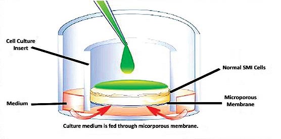
Figure 1: Schematic representation of the SI tissue model. SI epithelial cells are seeded onto a collagenfibroblast gel in cell culture inserts and air-lifted so that the apical tissue surface is exposed to the atmosphere. The tissue is fed by medium diffusing through the microporous membrane on the bottom of the cell culture insert. Test articles can be exposed to the tissue by topically application or by adding them to the culture medium (systemic exposure.)
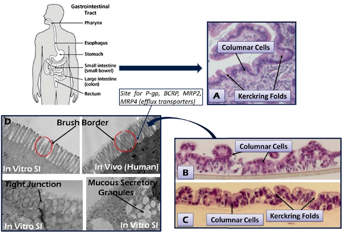
Figure 2: H&E staining showing the: (A) epithelium of explant small intestinal tissue, (B) partial thickness (PT), and (C) full-thickness (FT) small intestine (SI) tissue model. Micrographs of brush border membranes, tight junction formation and mucous secretory granules are also shown.
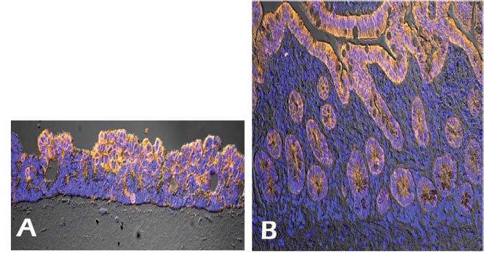
Figure 3: Immunohistochemistry showing cytokeratin 8 (CK8) expression (orange) by the reconstructed SI tissue model (A) and explant of small intestinal tissue (B).
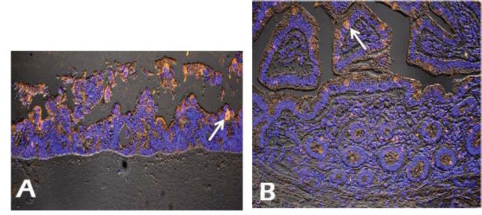
Figure 4: Immunohistochemistry showing MUC2 expression (arrow) by the reconstructed SI tissue model (A) and explant of small intestinal tissue (B).
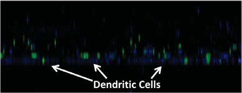
Figure 5: Confocal microscopic observation showing incorporation of dendritic cells (DC, green) into the SI tissue model. DC were stained using CellTrace –CFSE staining. Blue indicates nuclear staining.
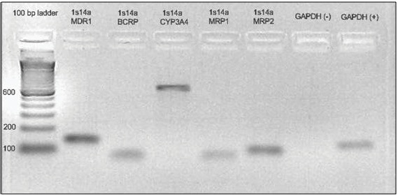
Figure 6: RT-PCR showing gene expression of the pglycoprotein
(MDR1), the metabolic enzyme CYP3A4, other drug transporters (multidrugresistance
proteins (MRP-1 and MRP-2), and the breast cancer resistance protein (BCRP) in the in
vitro reconstructed human SI tissue model.
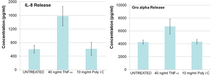
Figure 7: Cytokine analysis following acute exposure (24 hr) of SI tissue to TNF-α and
GRO-α. Culture supernatants were collected and analyzed for released cytokines/chemokines using Bio-Plex ELISA.

포스터 요청은 이곳에서 해주세요.