EpiCorneal Tissue Model

Features
EpiCorneal Tissue Model
- Corneal Three Dimensional Structure with Tight Junctions
- Constructed with Normal Human Corneal Epithelial Cell
- Native Corneal Morphology, Physiology & Barrier
- Quantifiable, Objective Endpoints
- Topical Testing of OphthalmicCost Effective Alternative to Animal Testing
General MatTek Tissue Features
- Unsurpassed Long-Term Tissue Reproducibility
- 3-Dimensional, Highly Differentiated Tissue
- Metabolically, Mitotically Active
- Produced from Normal (Non-Transformed) Human Cells - Ideal for Genomics Studies
- Produced in Easily Handled Cell Culture Inserts
- Grown in Completely Serum-Free Media System
- Quantifiable, Objective Test Endpoints
- Cost Effective Alternative to Animal and Human Clinical Testing
Applications
• Drug Delivery ( Drug Permeability )
• Infection
• Eye-related Bioavailability Assessment
• Inflammation
• Ophthalmic Product Testing
Data Sheet
MatTek’s EpiCorneal ocular tissue model consists of normal human Corneal epithelial cells which have been cultured to form a stratified, squamous epithelium that closely parallels normal human corneal tissue. The corneal cells, which are cultured on specially prepared cell culture inserts using serum free medium, differentiate to form a multi-layered structure containing tight junctions and express cornea-specific drug transporters and enzymes. Water soluble, non-water soluble and neat test materials can be directly applied to EpiCorneal tissue model.
The EpiCorneal tissue model exhibits in vivo-like morphological and growth characteristics which are uniform and highly reproducible. EpiCorneal consists of highly organized basal cells which progressively flatten out as the apical surface of the tissue is approached, analogous to the normal in vivo corneal epithelium. EpiCorneal is mitotically and metabolically active and releases many of the pro-inflammatory agents (cytokines) known to be important in ocular irritation and inflammation.
Various pharmacological and ophthalmological laboratories are actively seeking alternatives to isolated rabbit corneal permeability testing. Our data indicates that EpiCorneal can effectively provide a human-relevant, in vitro means to assess ocular irritation, drug delivery, endomicrobial and other infections, as well as wound healing and tissue regeneration studies. EpiCorneal is produced utilizing Good Manufacturing Practice (GMP) procedures making them highly reproducible.
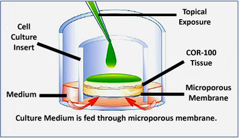
Figure 1: Schematic Representation of a topical exposure using EpiCorneal. Grown in tissue culture inserts at the air-liquid interface, the apical surface of EpiCorneal is exposed to air.
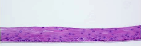
Figure 2: H&E stained histological cross-sections of EpiCorneal. Tissue structure closely parallels human corneal epithelium and includes the basal cell layer, wing cell layers and squamous cell layers. 10X magnification.
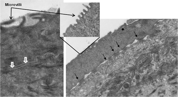
Figure 3: Ultrastructural features of EpiCorneal. The apical cell layer of EpiCorneal exhibits microvilli (open arrows), tight junctions (white arrows), and desmosomes (closed arrows). Tight junctions (white arrows) and desmosomes (closed arrow) are observed at the junction of adjacent epithelial cells.
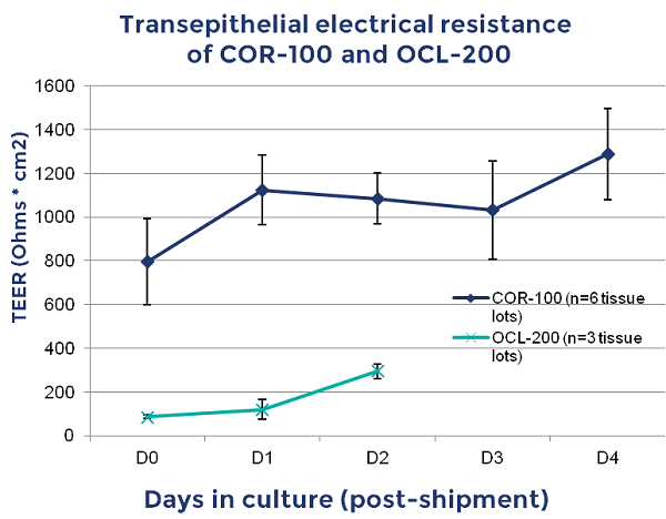
Figure 4: Transepithlial Electrical Resistance (TEER) of EpiCorneal (COR-100, comprised of corneal epithlial cells) and EpiOcular (OCL-200, comprised of keratinocytes). Note high TEER of COR-100 (avg.> 600 Ω*cm2) when compared to OCL-200 (~200Ω*cm2).
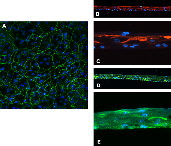
Figure 5: Expression of cornea-specific markers by EpiCorneal. Immunohistochemical staining for: A. Tight junction protein ZO-1 (green, top view), B. and C. Cytokeratin 3/Cytokeratin 12 (red), D. and E. Aldehyde Dehydrogenase-3 (ALDH-3A1) (green, cross section). Blue - DAPI (nuclei).
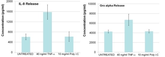 Figure 6: Gene expression of drug transporters and cornea-specific markers in EpiCorneal.